IBDP> CORE TOPICS> TOPIC 6 HUMAN HEALTH AND PHYSIOLOGY> 6.1 DIGESTION AND ABSORPTION
OPTION-TOPIC D- HUMAN PHYSIOLOGY> ; D-2 DIGESTION
LESSON PLAN
Animations and links
Pre-knowledge assessment
DAY 1
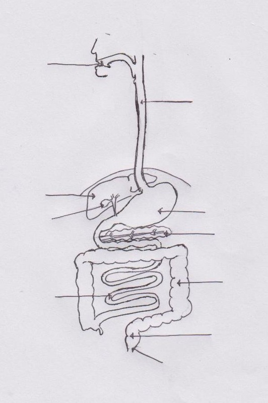
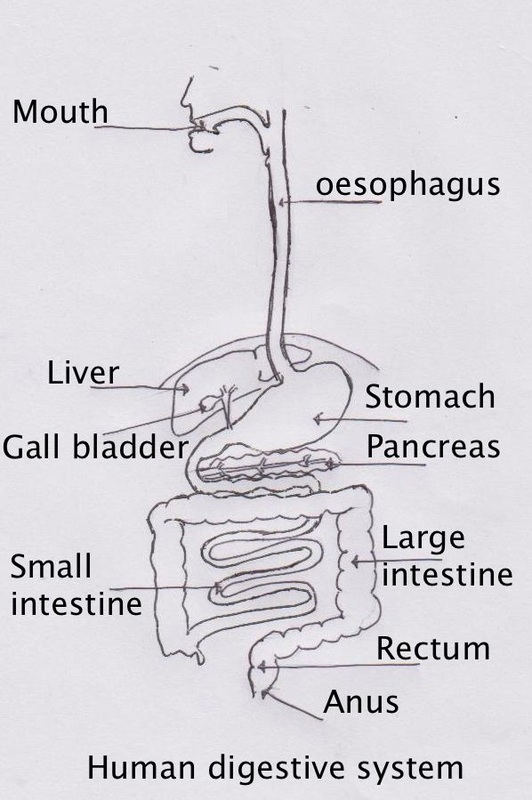
6.1.S1: Production of an annotated diagram of the digestive system
Points for drawing (4 marks)
|
|
DAY 2
D.2.U2: Exocrine glands secrete to the surface of the body or the lumen of the gut
D.2.U4: Acid conditions in the stomach favour some hydrolysis reactions and help to control pathogens in ingested food
D.2.S1: Identification of exocrine gland cells that secrete digestive juices and villus epithelium cells that absorb digested foods from electron micrographs
D.2.NOS: Serendipity and scientific discoveries—the role of gastric acid in digestion was established by William Beaumont while observing the process of digestion in an open wound caused by gunshot
|
D.2.U2: Exocrine glands secrete to the surface of the body or the lumen of the gut
|
D.2.U4: Acid conditions in the stomach favour some hydrolysis reactions and help to control pathogens in ingested food
|
|
|
|
D.2.S1: Identification of exocrine gland cells that secrete digestive juices and villus epithelium cells that absorb digested foods from electron micrographs
DAY 3
D.2.U1: Nervous and hormonal mechanisms control the secretion of digestive juices
D.2.U3: The volume and content of gastric secretions are controlled by nervous and hormonal mechanisms
D.2.A3: Helicobacter pylori infection as a cause of stomach ulcers
D.2.A1: The reduction of stomach acid secretion by proton pump inhibitor drugs
6.1.U2: The pancreas secretes enzymes into the lumen of the small intestine (amylase, lipase, endopeptidase)
|
D.2.U1: Nervous and hormonal mechanisms control the secretion of digestive juices
|
D.2.U3: The volume and content of gastric secretions are controlled by nervous and hormonal mechanisms
|
|
D.2.Application 3: Helicobacter pylori infection as a cause of stomach ulcers
|
D.2.Application 1:The reduction of stomach acid secretion by proton pump inhibitor drugs
|
DAY 4
6.1.U3: Enzymes digest most macromolecules in food into monomers in the small intestine
6.1.U1: The contraction of circular and longitudinal muscle of the small intestine mixes the food with enzymes and moves it along the gut
6.1.U5: Villi absorb monomers formed by digestion as well as mineral ions and vitamins
6.1.U4: Villi increase the surface area of epithelium over which absorption is carried out
6.1.S2: Identification of tissue layers in transverse sections of the small intestine viewed with a microscope or in a micrograph
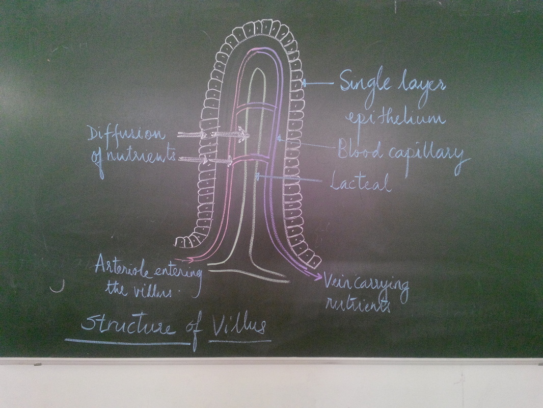
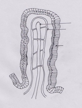
The structure of the villus is very specific. Firstly there is a great number of them so this increases the surface area for absorption in the small intestine. In addition the villi also have their own projections which are called microvilli. The many microvilli increase the surface area for absorption further. These microvilli have protein channels and pumps in their membranes to allow the rapid absorption of food by facilitated diffusion and active transport.
Adaptations of the villus
- There is a great number of VILLI so this increases the surface area for absorption in the small intestine.
- In addition the villi also have their own projections which are called microvilli. The many microvilli increase the surface area for absorption further.
- The villi contains an epithelial layer which is only one cell layer thick so that food can pass through easily and be absorbed quickly.
- The blood capillaries in the villus are very closely associated with the epithelium so that the distance for the diffusion of the food molecules is small.
- This thin layer of cells contains mitochondria to provide the ATP needed for the active transport of certain food molecules.
- There is a lacteal branch at the centre of the villus which carries away fats after absorption.
|
This video will help you understand the folds, Villus and microvilli in the small intestine
|
6.1.U4: Villi increase the surface area of epithelium over which absorption is carried out
|
DAY 5
D.2.U5: The structure of cells of the epithelium of the villi is adapted to the absorption of food
6.1.U6: Different methods of membrane transport are required to absorb different nutrients
6.1.U6: Different methods of membrane transport are required to absorb different nutrients
DAY 6 : LAB SESSION
6.1.A1: Processes occurring in the small intestine that results in the digestion of starch and transport of the products of digestion to the liver
6.1.A2: Use of dialysis tubing to model absorption of digested food in the intestine
6.1.A2: Use of dialysis tubing to model absorption of digested food in the intestine
DAY 7
6.1.NOS: Use models as representations of the real world-dialysis tubing can be used to model absorption in the intestine
D.2.U7: Materials not absorbed are egested
D.2.U6: The rate of transit of materials through the large intestine is positively correlated with their fibre content
D.2.A2: Dehydration due to cholera toxin
D.2.U7: Materials not absorbed are egested
D.2.U6: The rate of transit of materials through the large intestine is positively correlated with their fibre content
D.2.A2: Dehydration due to cholera toxin
TOK : Models are often used to better understand nature - use of visking tubes for modelling digestion.
Skills- Draw and label the diagrams
Digestive system
|
|
|
Structure of villus
|
|
ACTIVITY
Nov - 2020. A movie on working of digestive system
Students work in groups to create a story board/game of the digestion and absorption
Digestive system model making- activity
References-
Damon, Alan et al, Higher level Biology for the IB diploma. Pearson Baccalaureate
Clegg, CJ, Biology for the IB diploma. London: Hodder Murrray, 2007, 978-0340926529
Taylor, Stephan, Science Video resources Wordpress,
Burell, John. Click 4Biology(online)
Chris Paine- https://www.bioknowledgy.info/
Brent Cornell- https://ib.bioninja.com.au/
Gretel von Bargen- https://www.biologyforlife.com/syllabus.html
All pictures have been downloaded from Google images for educational purpose only.