IBDP> CORE TOPICS> TOPIC 6 HUMAN HEALTH AND PHYSIOLOGY>
6.4 GAS EXCHANGE
ASSESSMENT STATEMENTS6.4.1 Ventilation, gas exchange and cell respiration.
6.4.2 The need for a ventilation system. 6.4.3 Features of the alveoli. 6.4.4 Structure of the ventilation system. 6.4.5 Mechanism of ventilation. |
HELPFUL LINKS |
Introductory video
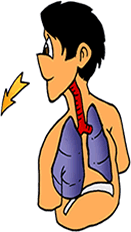
6.4.1 Distinguish between ventilation, gas exchange and cell respiration.
|
All organisms need oxygen. Oxygen is required during cellular respiration. This is a biochemical pathway that involves the sequential breakdown of chemical bonds of the glucose molecule. This process requires oxygen and gives off carbon di oxide.
our lungs with the heart and blood vessels ensure that all the cells receive this supply of oxygen. Throughout our lives we continuously fill our lungs with air and then breathe it out. This process of taking in air and breathing out of air is called ventilation. The oxygen in the lungs diffuse into the bloodstream and the carbon di oxide in the blood stream diffuses into the lungs. Each breath we take replenishes the supply of gases within the lung tissues so that diffusion continues. This process is called gas exchange. Respiration is the process of breakdown of glucose in the cell for release of energy in the form of ATP. |
|
Outline the process of gas exchange-
Gas exchange occurs at two locations-
1. In the lungs- Oxygen move out from the air in the lungs to the RBC in the blood stream and carbon di oxide moves in the opposite direction.
2. In a capillary bed- Any place in the body where opposite gas exchange takes place. Oxygen diffuses into the body cell from the blood stream and carbon di oxide diffuses out of the body cell into the blood stream of the capillary bed.
Differentiate between ventilation and gas exchange
|
VENTILATION
|
GAS EXCHANGE
|
6.4.2 Explain the need for a ventilation system.
|
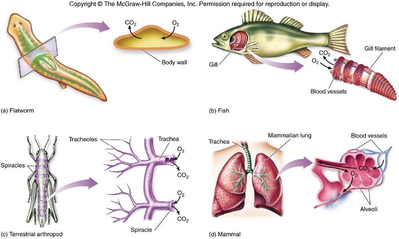
Small animals obtain oxygen by direct diffusion in the cells. Amoeba through the cell membrane, other organisms like planarians by outer membrane
In humans and other large animals skin is ineffective for ventilation. Humans are large and have a small ratio surface area : volume; so they need ventilation system. The ventilation system is needed for- 1. to increase surface area. 2. to maintain a concentration gradient in alveoli; as oxygen is used in respiration (and carbon dioxide is produced); The gaseous exchange occurs between air in alveoli and blood capillaries; alveoli have high ratio surface area : volume. This is to facilitate ventilation; or to bring in fresh air and remove stale air. |
|
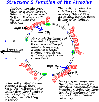
6.4.3 Features of the alveoli.
|
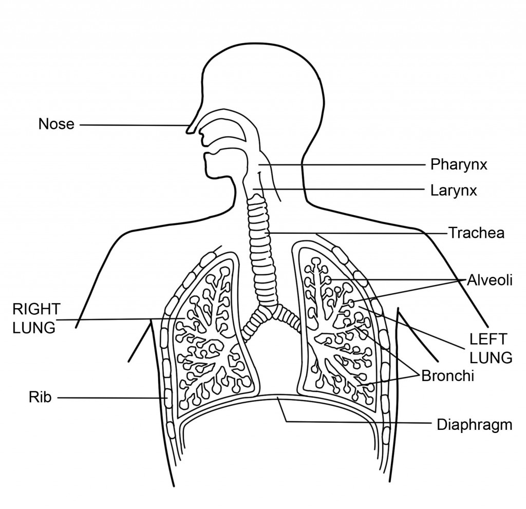
The air that is breathed through the nasal passage follows a pathway-
1. trachea 2. left and right primary bronchi 3. smaller branches of bronchi 4. very smaller branches- bronchiole 5. small air sacs- alveoli The alveoli are small sacs at the end of the smallest branches. There are approximately 300 million alveoli in each lung. The alveoli are surrounded by capillary bed. The blood entering the capillary bed is low in oxygen and high in carbon di oxide from the right ventricles. In the alveoli oxygen diffuses into the blood and carbon di oxide from the blood diffuses into the alveoli from the blood. The alveoli shows various adaptations- 1. Spherical shape- provides large surface area 2. Flattened, single cell thickness- facilitates easier diffusion of gases. 3. Moist inner lining of alveoli- allows efficient diffusion 4. Associated capillary bed nearby- the distance for diffusion is less. |
|
6.4.4 Structure of the ventilation system.
|
Points to be remembered for drawingTrachea to be shown with cartilage rings and labelled;
Bronchi and bronchioles shown and labelled; Two lungs shown and at least one of them labelled; Diaphragm shown with a dome shape and labelled; Ribs and intercostal muscles shown and labelled; Pharynx mouth and nasal passages shown |
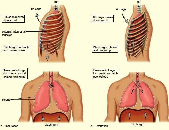
6.4.5 Mechanism of ventilation.
External intercostal muscles contract and internal intercostal muscles relax. This results in pulling the rib cage upwards. The diaphragm contracts and flattens which increase the volume of thoracic cavity. This reduces pressure; so air enters the lungs.
Internal intercostal muscles contract / external intercostal muscles relax; diaphragm relaxes; abdominal muscles push diaphragm upwards. This results in decrease in volume of thoracic cavity. Thus increasing the pressure; so air leaves the lungs.
Internal intercostal muscles contract / external intercostal muscles relax; diaphragm relaxes; abdominal muscles push diaphragm upwards. This results in decrease in volume of thoracic cavity. Thus increasing the pressure; so air leaves the lungs.
INSPIRATION
|
EXPIRATION
|
CLASS PRESENTATION
REFERENCES
Damon, Alan et al, Higher level Biology for the IB diploma. Pearson Baccalaureate
Clegg, CJ, Biology for the IB diploma. London: Hodder Murrray, 2007, 978-0340926529
Taylor, Stephan, Science Video resources Wordpress,
Burell, John. Click 4Biology(online)
All picture have been downloaded from Google images for educational purpose only
Clegg, CJ, Biology for the IB diploma. London: Hodder Murrray, 2007, 978-0340926529
Taylor, Stephan, Science Video resources Wordpress,
Burell, John. Click 4Biology(online)
All picture have been downloaded from Google images for educational purpose only