IBDP > CORE TOPICS > TOPIC 6. HUMAN HEALTH AND PHYSIOLOGY > 6.2 TRANSPORT SYSTEM
OPTION TOPIC D-2 THE HEART
LESSON PLAN
LINKS
DAY 1
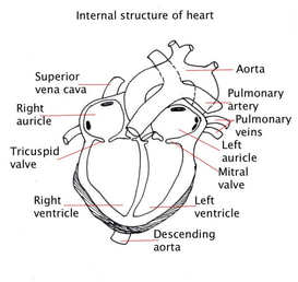
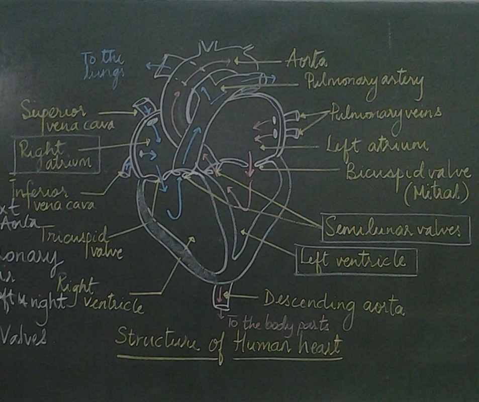
6.2.S2: Recognition of the chambers and valves of the heart and the blood vessels connected to it in dissected hearts or in diagrams of heart structure (Oxford Biology Course Companion page 295)
6.2.U7: There is a separate circulation for the lungs (Oxford Biology Course Companion page 295)
6.2.U6: Valves in veins and the heart ensure circulation of blood by preventing backflow (Oxford Biology Course Companion page 294).
6.2.U7: There is a separate circulation for the lungs (Oxford Biology Course Companion page 295)
6.2.U6: Valves in veins and the heart ensure circulation of blood by preventing backflow (Oxford Biology Course Companion page 294).
|
|
|
6.2.SKILL-2: Recognition of the chambers and valves of the heart and the blood vessels connected to it in dissected hearts or in diagrams of heart structure (Oxford Biology Course Companion page 295)
Heart has collecting chambers for the blood moving in from the veins.
These are thin walled muscular chambers called ATRIUM.
Heart has thick walled muscular chambers that builds up pressure to send the blood out with a force.
These chambers are called VENTRICLES.
The blood forced out of the heart completes a circuit to come back to the heart-
A large artery ---Smaller arteries-----Arteriole-----Capillary bed----Venule-----Larger Veins------Inferior vena cava that takes blood back to the heart.
The flow of blood is from atria to ventricle that is guarded by artrio-ventricular valves and from ventricle to the outside that is guarded by semilunar valves.
The walls of the ventricle are thicker than the atrium. The wall of the left ventricle is thicker than the right ventricle.
Points to remember for drawing
|
|
|
|
|
6.2.U7: There is a separate circulation for the lungs (Oxford Biology Course Companion page 295)
6.2.U6: Valves in veins and the heart ensure circulation of blood by preventing backflow (Oxford Biology Course Companion page 294).
6.2.U6: Valves in veins and the heart ensure circulation of blood by preventing backflow (Oxford Biology Course Companion page 294).
|
CORONARY CIRCULATION
The muscles of the heart do not receive nutrients and oxygen from the blood in the chambers heart. The muscles of the heart are supplied with a system of blood vessels. This is called as the coronary circulation. These are the branches of the aorta. The heart muscles receive nutrients and oxygen from these blood vessels. The right side of the heart sends blood to the lungs (the capillary bed). This is the PULMONARY CIRCULATION. The left side of the heart sends blood to all parts of the body organs (capillary bed). This is called SYSTEMIC CIRCULATION. |
|
DAY 2
D.4.NOS: Developments in scientific research followed improvements in apparatus or instrumentation—the invention of the stethoscope led to improved knowledge of the workings of the heart (Oxford Biology Course Companion page 687).
6.2.A3: Pressure changes in the left atrium, left ventricle and aorta during the cardiac cycle (Oxford Biology Course Companion page 300)
6.2.A3: Pressure changes in the left atrium, left ventricle and aorta during the cardiac cycle (Oxford Biology Course Companion page 300)
ASSIGNMENT: 6.2.A3: Pressure changes in the left atrium, left ventricle and aorta during the cardiac cycle
DAY 3
D.4.U1: Structure of cardiac muscle cells allows propagation of stimuli through the heart wall. (Oxford Biology Course Companion page 685).
6.2.U8: The heartbeat is initiated by a group of specialized muscle cells in the right atrium called the sinoatrial node (Oxford Biology Course Companion page 298).
6.2.U9: The sinoatrial node acts as a pacemaker(Oxford Biology Course Companion page 299).
6.2.U10: The sinoatrial node sends out an electrical signal that stimulates contraction as it is propagated through the walls of the atria and then the walls of the ventricles (Oxford Biology Course Companion page 299).
D.4.U2: Signals from the sinoatrial node that cause contraction cannot pass directly from atria to ventricles (Oxford Biology Course Companion page 685).
D.4.U3: There is a delay between the arrival and passing on of a stimulus at the atrioventricular node (Oxford Biology Course Companion page 686).
D.4.U4: This delay allows time for atrial systole before the atrioventricular valves close (Oxford Biology Course Companion page 687).
D.4.U5: Conducting fibres ensure coordinated contraction of the entire ventricle wall (Oxford Biology Course Companion page 687).
D.4.S3: Mapping of the cardiac cycle to a normal ECG trace (Oxford Biology Course Companion page 689)
ASSIGNMENT: D.4.S3: Mapping of the cardiac cycle to a normal ECG trace
ACTIVITIES - BLOOD CIRCULATION
ACTIVITY 1:
An RBC has a life span of 120 days. Consider your self to be an RBC trace alt east four possible circuits that you would complete during this time.
Find out the name of the artery that lead to different organs and the veins that carry them back to the heart.
You have to create a digital circuit diagram and submit it in the next slot.
Proper representation of pulmonary and systemic circulation 4
Names of all branches of arteries and veins 4 x 4
ACTIVITY 2:
THE DOUBLE CIRCULATION ACTIVITY
GROUP ACTIVITY-
You will work in pairs.
Each group is given one organ and a muscle. You have to trace the path travelled by an RBC to both these parts of the body.
The organ is common for all groups- LIVER
GROUP 1- MUSCLE TIBIALIS
GROUP 2- MUSCLE ULNARIS
GROUP 3- MUSCLE FRONTALIS
You have to prepare a flow chart of the double circulation.
Draw the main organs and the blood vessels and colour them accordingly.
Mention the names of branch of each artery and vein leading to the organ and muscle.
You will be presenting this in the next slot.
You will be graded on your presentation
Proper representation of pulmonary and systemic circulation 4
Names of all branches of arteries and veins 4
Neat and clear representation 2
Proper explanation 3
Team work 2
STUDENTS WILL COMPLETE THIS AS HOMEWORK ASSIGNMENT
DAY 4
D.4.A1: Use of artificial pacemakers to regulate the heart rate (Oxford Biology Course Companion page 689).
D.4.A2: Use of defibrillation to treat life-threatening cardiac conditions (Oxford Biology Course Companion page 690).
6.2.U11: The heart rate can be increased or decreased by impulses brought to the heart through two nerves from the medulla of the brain (Oxford Biology Course Companion page 301).
6.2.U12: Epinephrine increases the heart rate to prepare for vigorous physical activity(Oxford Biology Course Companion page 302).
D.4.S1: Measurement and interpretation of the heart rate under different conditions (Oxford Biology Course Companion page 688).
6.2.A1: William Harvey’s discovery of the circulation of the blood with the heart acting as the pump (Oxford Biology Course Companion page 290).
6.2.NOS: Theories are regarded as uncertain- William Harvey overturned theories developed by the ancient Greek philosophy Galen on movement of blood in the body (Oxford Biology Course Companion page 290).
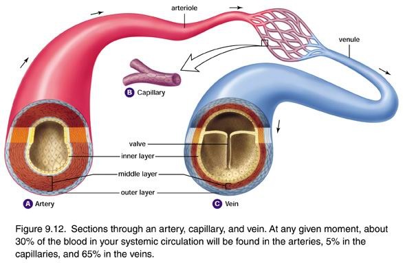
6.2.U1: Arteries convey blood at high pressure from the ventricles to the tissues of the body (Oxford Biology Course Companion page 291).
6.2.U2: Arteries have muscle cells and elastic fibres in their walls (Oxford Biology Course Companion page 291).
6.2.U3: The muscle and elastic fibres assist in maintaining blood pressure between pump cycles (Oxford Biology Course Companion page 292).
6.2.U4: Blood flows through tissues in capillaries. Capillaries have permeable walls that allow exchange of materials between cells in the tissue and the blood in the capillary (Oxford Biology Course Companion page 293).
6.2.U5: Veins collect blood at low pressure from the tissues of the body and return it to the atria of the heart (Oxford Biology Course Companion page 293).
6.2.A2: Causes and consequences of occlusion of the coronary arteries (Oxford Biology Course Companion page 297).
D.4.A3: Causes and consequences of hypertension and thrombosis (Oxford Biology Course Companion page 690).
D.4.S4: Analysis of epidemiological data relating to the incidence of coronary heart disease
(Oxford Biology Course Companion page 692).
D.4.A2: Use of defibrillation to treat life-threatening cardiac conditions (Oxford Biology Course Companion page 690).
6.2.U11: The heart rate can be increased or decreased by impulses brought to the heart through two nerves from the medulla of the brain (Oxford Biology Course Companion page 301).
6.2.U12: Epinephrine increases the heart rate to prepare for vigorous physical activity(Oxford Biology Course Companion page 302).
D.4.S1: Measurement and interpretation of the heart rate under different conditions (Oxford Biology Course Companion page 688).
6.2.A1: William Harvey’s discovery of the circulation of the blood with the heart acting as the pump (Oxford Biology Course Companion page 290).
6.2.NOS: Theories are regarded as uncertain- William Harvey overturned theories developed by the ancient Greek philosophy Galen on movement of blood in the body (Oxford Biology Course Companion page 290).
6.2.U1: Arteries convey blood at high pressure from the ventricles to the tissues of the body (Oxford Biology Course Companion page 291).
6.2.U2: Arteries have muscle cells and elastic fibres in their walls (Oxford Biology Course Companion page 291).
6.2.U3: The muscle and elastic fibres assist in maintaining blood pressure between pump cycles (Oxford Biology Course Companion page 292).
6.2.U4: Blood flows through tissues in capillaries. Capillaries have permeable walls that allow exchange of materials between cells in the tissue and the blood in the capillary (Oxford Biology Course Companion page 293).
6.2.U5: Veins collect blood at low pressure from the tissues of the body and return it to the atria of the heart (Oxford Biology Course Companion page 293).
6.2.A2: Causes and consequences of occlusion of the coronary arteries (Oxford Biology Course Companion page 297).
D.4.A3: Causes and consequences of hypertension and thrombosis (Oxford Biology Course Companion page 690).
D.4.S4: Analysis of epidemiological data relating to the incidence of coronary heart disease
(Oxford Biology Course Companion page 692).
LAB WORK 6.2.S1: Identification of the blood vessels as arteries, capillaries or veins from the structure of their walls (Oxford Biology Course Companion page 294).
Observe the permanent slides of artery and vein.
Try to identify the layers and write down your observation
Observe the permanent slide of blood smear.
Try to identify the different types of blood cells and their features and write down your observations with drawings.
Try to identify the layers and write down your observation
Observe the permanent slide of blood smear.
Try to identify the different types of blood cells and their features and write down your observations with drawings.
LAB WORK: D.4.S2: Interpretation of systolic and diastolic blood pressure measurements (Oxford Biology Course Companion page 691).
Structure and function of arteries, capillaries and veins.
|
The blood vessels are -
Arteries- that carry blood away from the heart to tissues; Arteries have thick walls to withstand high pressure. This prevents bursting of arteries due to high pressure. Arteries have muscle fibres to even out blood flow and elastic fibres to allow artery wall to stretch/recoil. Veins- veins carry blood back to the heart from the tissues. Veins have thinner walls because the pressure is low. Veins have fewer muscle / elastic fibres because there is no pulse. Veins have valves to prevent backflow; Capillaries – These are the minute vessels that allow exchange of O2 / CO2 / nutrients / waste products from tissues. Capillaries have a thin wall to allow (rapid) diffusion. Capillaries have porous walls to allow tissue fluid to leave. Capillaries are narrow so can penetrate all parts of tissues, bigger total surface area. |
|
References-
Damon, Alan et al, Higher level Biology for the IB diploma. Pearson Baccalaureate
Clegg, CJ, Biology for the IB diploma. London: Hodder Murrray, 2007, 978-0340926529
Taylor, Stephan, Science Video resources Wordpress,
Burell, John. Click 4Biology(online)
Chris Paine- https://www.bioknowledgy.info/
Brent Cornell- https://ib.bioninja.com.au/
Gretel von Bargen- https://www.biologyforlife.com/syllabus.html
All pictures have been downloaded from Google images for educational purpose only.